Throughout my residency I have found these three images to have particular significance
Sketch 'n Scrubs
Herein is a sharing of my creative process and experience in North Bay as Artist in Residence within the unique setting of a hospital through its ArtsHealth North Residency Program. The residency is from September 1 to November 11 2011
Wednesday, November 16, 2011
A small grouping of recurring motifs
Around the end of October I found discarded copies of the architectural drawings and floor plans of the health centre . I got in touch with the architect for permission to use as a background to some of my drawings. Here are the results of the combination.
Completing my work here
Apart from the set of threes I also completed these 2 drawings in graphite and charcoal
A closer view of the twisters. I was wanting a form to echo that of the roots but with an opposite actionSaturday, October 29, 2011
Unique things to see and do in North Bay
Edible Plant Herb Walk:
A local resident named Carlos in North Bay occasionally leads small groups in the neighboring woods to identify many wild edible plants. He explained how they can be prepared for food. Notebooks in hand we enjoyed the hike and the vast knowledge Carlos shared with us.
Home Concerts:
Piebird Farms Bed & Breakfast offer concerts, dinners and educational workshops on growing and preparing foods and on wellness with herbs. I enjoyed a concert by Faye Blais who played folk music in the living room while we sat cozy on an autumn night. It is seriously a very unique and wonderful place to experience. See links to things mentioned on the side bar of this blog.
Local Symphony:
I took in a concert of the North Bay Symphony at the Capitol Theatre. It's a beautiful space and I really enjoyed the symphony's program. I found the pieces chosen accessible and what the conductor shared about them made it all more informative and delightful. I also got to enjoy the Silver Birch Quartet and oboist Erin Brophey.
Delaney Bay Cheese Market:
A seriously wide selection of artisan and farmstead cheeses with wild game charcuterie. There are also great balsamic vinegar, jellies, chutneys and my favorite compote of figs with Porto. The owner Lonnie Reed made some food and wine pairing suggestions that were amazing. You foodies out there will need to check this place out.
Local Farmer's Market:
It starts May long weekend every Saturday morning right up to Thanksgiving weekend. This is where I got my weekly groceries and met some of the local folks. People around here go to say hi to friends even if they're not buying. The market's got great energy and I have made some connections with some of the local farmers. Check out Field Good Farms in the side links to things mentioned.
Gallery Openings and Events:
There are many artists living in North Bay who are very involved with the galleries and associations. I find refreshing in small regions that few divisions exist between the public, artist run and commercial galleries. On October 14th there is a wonderful gallery hop night and opportunity to meet and chat with the local scene. This is an image of Amanda Burk's work exhibited in the Kennedy Gallery which is the public gallery here in North Bay. Amanda has recently opened the Line Gallery which is dedicated to exhibiting and documenting contemporary Canadian drawing. See links to things mentioned for these galleries.
Cool local shops:
My favorite local shops are Farm on Main Street seen in this image and Déjà Vu a savvy recently opened retro and antique home accessories boutique on Oak Street.
Saturday, October 22, 2011
Tuesday, October 18, 2011
Visit of the Diagnostic Imaging Department
Taken in 1940 by an unknown photographer this photo of the male technician and female patient was used in the day to demonstrate the "myth" (my quotations) about exposure to radiation during the x-ray procedure (courtesy of the US National Cancer Institute).
Arlie Hoffman worked many years as Technical Director of radiology and is now dedicated full time to his painting. Upon meeting me and seeing my current fascination with x-ray images he generously offered to set-up a tour for me of the Diagnostic Imaging Department and contacted Suzanne Mitchell (right) who is one of the Charge Technologists. Between the two is Chantal Speirs the Studio Technician at ArtsHealth North Residency and go-to person for all things art in the hospital (i.e. permanent art collection and residency program).
Images were once limited to x-rays so most hospitals referred to their Diagnostic Imaging Department as their X-ray Department. Now images include MRI (Magnetic Resonance Imaging), CT (Computed Tomography), Nuclear Medicine (injectable radioisotopes) and Ultra Sound (Sonography Imaging). Suzanne walked me through the various rooms and explained some of the history of these technologies.
This is a typical rotating anode x-ray tube that actually produces x-rays in machines today. I worried it might be radioactive but Sue explained that the tube produces radiation when activated but does not store it hence being safe to touch. Like any vacuum tube, the little disc-shape part in the middle is the rotating anode. The cathode in front of it (left part of the tube) emits photons (energy released by electrons) which collide on the anode plate and bounce off into the tube. The x-ray spectrum depends on the anode material and accelerating voltage producing the rotation. The anode plate usually made of tungsten or molybdenum in this tube looks porous due to the wear and tear caused by photons hitting its surface. Although not visible here, there is a tiny hole in the glass located between the anode and cathode called a window. When the tube is inserted in the machine it is surrounded in an oil bath encased in lead to avoid radiation leakage.
This lead cone beam limiting device is an antique salvaged from the old hospital site. The narrow end of the cone shown here was placed up against the glass anode tube's window mentioned earlier.
The wider part of the cone was placed above the body of the person diagnosed. The radio active photons bouncing off the anode plate would escape the glass enclosure through the window and move down this tube towards the wider part of the cone. The current beam limiting devices automatically cone to the area of interest. Please visit the links section on this blog if you want to learn more on the subject of x-rays.
I first got to see the equipment for MRI (Magnetic Resonance Imaging). Unlike a CT it works with magnetic fields and radio waves instead of x-rays to create images. The MRI is dependent on the movement of the hydrogen atoms in the body. These atoms usually go in different directions but when placed in the magnetic field created by this machine, the hydrogen atoms in the body line up in the direction of the field. When the hydrogen atoms return to their original state the movement is detected and retained as an image on a computer screen. This diagnostic tool is best for soft tissue delineation because the tissue is abundant with water. The software for these devices is absolutely astounding in its sophistication and precision. I just kept staring at the screen in total awe.
To protect the privacy of patients I could not take pictures of the screen. I have found on the web a very similar image to what I saw. The difference is that this image here is static while what I was looking at was constantly shifting. The Technologist could stop the movement at any time to analyse and flip to various plane views of the same spot of the body.
This is a CT (Computed Tomography) machine. Although it looks similar to the MRI it works very differently and is basically density dependent. A CT is a very comprehensive x-ray. The x-ray detector revolves around the body and varies the intensity of rays in order to scan each type of tissue. Depending on the pathology and tissue affected it may be better to go with one type of scan over an other. Unfortunately given the CT is so comprehensive it means more radiation exposure for the patient. Doctors decide whether the diagnostic information outweighs the radiation exposure for the patient and each case considered very carefully. Sue explained that CT is a great tool for virtual autopsy as well because the corpse would not have to be altered by manual autopsy process.
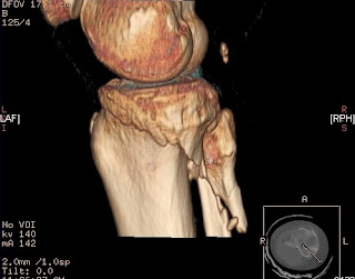
The image results of the CT is what amazed me the most of the entire tour. I was mesmerized by all the planes and angle views we could observe of one specific part of the body. One of the Technologists showed me a knee image and made the image flip around 180 degrees. Then he showed me how he could have the image remove the femur and angle the view from above the tibia and fibula as though we were in the place of the femur. This totally flipped me out! Although there are programs for architecture and animation that allow 3D renderings of something imagined the difference with a CT is that the image is from inside the actual body.
The rotations and unique points of views allow the Technologists to see injuries with very precise information not possible in the past without operating or opening the body (and even then the other tissues and blood could easily get in the way). I pondered this wonder all weekend and kept reviewing the image in my mind. I liken this view of the knees to seed pods.
Sunday, October 16, 2011
Making new grounds
I decided to paste the drawings together and create irregular shapes with the paper. I then covered these pasted drawings with a primer. The original bone drawings are mostly gone now but they serve as a ground for new drawings to emerge.
These are some works in progress so they may change significantly from now until they are completed.
I decided to keep 2 of the bone drawings. I cut parts of an earlier larger drawing I'd done (see earlier posts) and covered the whole thing with primer as well. I then pasted the bone drawings onto them to build the drawings from there.
Subscribe to:
Posts (Atom)


